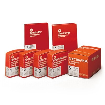
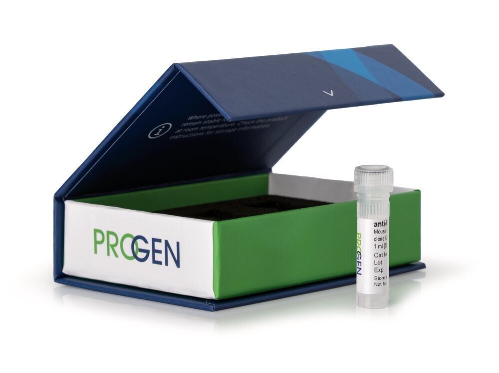
anti-Desmocollin 3 monoclonal de souris, Dsc3-U114, 1 ml
This antibody portfolio includes a wide range of tissue- & tumor-specific primary antibodies for basic research, product development and analysis of a variety of biological questions. The monoclonal antibodies are perfectly applicable for all cell or tissue biology studies and for the analysis of predictive markers in research.
- High epitope affinity
- Highly specific detection of the target protein
- Unconjugated
- Suitable for e.g. western blot, immunoprecipitation, ELISA & immunofluorescence
anti-Desmocollin 3 mouse monoclonal, Dsc3-U114 50 μg/ml, monoclonal, affinity purified
The mouse monoclonal antibody localizes Dsc3 present in all living epidermal layers, in glandular duct cells, in basal matrix cells and outer root sheath of hair follicles, in basal and suprabasal cell layers of stratified epithelia (e.g. vagina, tongue, esophagus), in basal layer of bladder urothelium and complex epithelium of trachea, in thymic reticulum cellsAll simple epithelia are negative. Intercalated disks of myocardium are negative.
Polypeptide reacting: 109 kDa and 100 kDa polypeptides (desmocollin 3 splice isoforms at pI 5.2) from human epidermal desmosomes.
Reactivity on cultured cell lines: most cultured cell lines derived from stratified squamous epithelia or squamous cell carcinomas are positive (e.g. A-431, HaCaT).
Host: mouse
Antibody type: monoclonal
Isotype: IgG1
Clone: Dsc3-U114
Immunogen: synthetic peptide corresponding to a sequence present on the extracellular anchor domain of human desmocollin 3 (VHGAPFYFSLPNTSPEISRLWSLTKVC)
UniprotID: Q14574 (human), P55292 (mouse), D3ZZI6 (rat)
Synonym: desmocollin-3, cadherin family member 3, desmocollin-4, HT-CP, DSC3, CDHF3, DSC4
Conjugate: unconjugated
Purification: affinity chromatography
Intended use: research use only
Application: IHC, WB
Reactivity: human, mouse, rat
No reactivity: bovine
Immunohistochemistry (IHC) - frozen: 1:10-1:100 (0.5-5 μg/ml, for better resolution preincubation (directly after fixation) with 0.05-0.2% Triton X-100, for 5-10 min, depending on tissue type, is recommended)
Immunohistochemistry (IHC) - paraffin: 1:10-1:100 (0.5-5 μg/ml, microwave treatment recommended)
Western Blot (WB): assay dependent


![Ethyl [(2-methoxyphenyl)carbamoyl]formate, 97%, 2g Ethyl [(2-methoxyphenyl)carbamoyl]formate, 97%, 2g](https://d2j6dbq0eux0bg.cloudfront.net/images/88473019/4769720813.png)
![(S)-2-[(Diphenylphosphino)methyl]pyrrolidine, 97%, 250mg (S)-2-[(Diphenylphosphino)methyl]pyrrolidine, 97%, 250mg](https://d2j6dbq0eux0bg.cloudfront.net/images/88473019/4763218337.png)
![N-(4-Chloro-7H-pyrrolo[2,3-d]pyrimidin-2-yl)-2,2-dimethylpropionamide, 95.0%, 1g N-(4-Chloro-7H-pyrrolo[2,3-d]pyrimidin-2-yl)-2,2-dimethylpropionamide, 95.0%, 1g](https://d2j6dbq0eux0bg.cloudfront.net/images/88473019/4780605142.png)
